
Why is adenomyosis diagnosis so difficult? Why is MRI more accurate than ultrasound in diagnosing adenomyosis?
Why ultrasound often missed the diagnosis of adenomyosis?
Women with adenomyosis typically complain of heavy menstrual bleeding (HMB) and severe period pain. These however are very common but non-specific symptoms. The uterus can be enlarged and sore when touched. Transvaginal ultrasound (TVUS) can suggest the diagnosis. However, the signs on ultrasound are often subtle, sonographers may need to be prompted to look for the subtle signs of adenomyosis. MRI is more accurate than TVUS, especially when the adenomyosis is superficial or diffuse, and when coexisting fibroids maybe confused with focal adenomyosis.
There is a wide variety of adenomyosis. This probably why adenomhyosis can be difficult to diagnose with ultrasound. MRI can see these changes for more accurately.
Adenomyosis is not a single disease. There are different types of adenomyosis. Adenomyosis is a spectrum of disease, from superficial to deep, from focal to diffuse. See this proposed classification of adenomyosis illustrated (by Bazot).
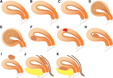
Classification of adenomyosis
A, B and C: superficial adenomyosis; D and E: deep diffuse; F: focal adenomyosis (AKA adenomyoma); D: cystic focal adenomyosis; H: Inside uterine cavity; I, J and K: outside the uterus (can be seen as endometriosis on laparoscopy)
Seeing this variety of adenomyosis, it is not hard to understand why ultrasound is struggling to make the accurate diagnosis of adenomyosis. It is the limitation of the ultrasound technology. In fact 71% of the ultrasound in our region missed the diagnosis of adenomyosis.
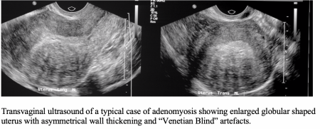
When adenomyosis is diffuse and superficial, the signs on ultrasound can be subtle. When it is more extensive, the uterus is globular in shape, often with one wall thicker than the other, and “Venetian blinds” maybe present. When adenomyosis is focal, it is called adenomyoma, which is often mistaken as fibroid on ultrasound. MRI is far more accurate than ultrasound in picking up subtle adenomyosis and when fibroids are also present.
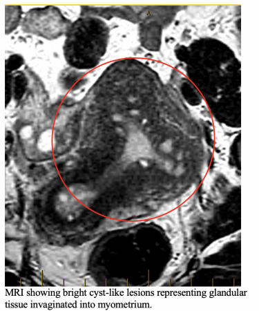
MRI detect bright signal glands (adeno) within muscle wall ( myosis), as well the reactive muscle cell changes ( seen as thickening of junctional zone).
Check out our Case Studies. In the first 2 cases ultrasound missed adenomyosis; in the other 2 cases ultrasound mistaken focal adenomyosis as fibroids!
